
Noses and Toes Gone Wrong
Numerous conditions that can affect your dog, from nose-tip to toenails.
Article by CJ Puotinen and Mary Straus, published in the Whole Dog Journal, August 2011
 Contents
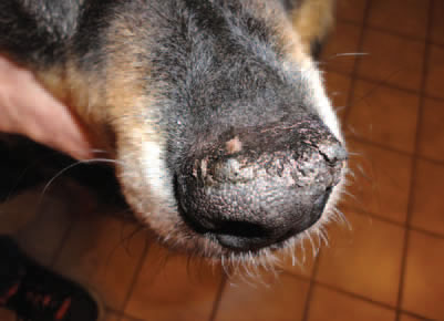
The photo above is of a dog with hyperkeratosis. Milo’s nose kept developing rough spots and cracking, causing the English Shepherd extreme pain. Photo courtesy of Katie Palmer.
Yikes! What happened to Fido’s nose? And what’s wrong with Fluffy’s paw pads? The possibilities are many, and a surprising number of nose and paw pad problems are related. Because illnesses in this category often have similar or identical symptoms, a veterinarian’s diagnosis can be important. The following overview will help you identify, prevent, or treat these disorders.
Update January 2020: See Watch for these red flags in your veterinary derm exams, especially the sections on "Appearance of Nasal Planum" and Paws," for some details about how to differentiate between various causes of abnormalities.
Pigment problems
The most frequently asked questions about dogs’ noses concern color. Dogs have black or dark noses and paw pads because of melanin, a pigment that darkens skin. When melanin production slows or stops, the skin lightens uniformly or in patches.
Nasal hypopigmentation, also known as nasal depigmentation, is most commonly seen in the Golden Retriever, yellow Labrador Retriever, white German Shepherd, Poodle, Doberman Pinscher, Irish Setter, Pointer, Samoyed, Siberian Husky, Malamute, Afghan Hound, and Bernese Mountain Dog. Nose color is normal at birth but gradually fades to a light brown or whitish color.
Considered harmless, hypopigmentation does not make the nose more sun-sensitive and does not require treatment. However, this cosmetic imperfection matters to breeders because the loss of nose pigment is a conformation fault in the show ring.
Vitiligo (pronounced vit-ill-EYE-go) creates white spots of varying size and location on the skin when pigment cells, or melanocytes, are destroyed, preventing melanin production. Dogs with this immune system disorder develop white spots on the nasal planum (the hairless, leathery part of the nose), muzzle, and inner lining of the cheeks and lips, as well as patches of white hair and scattered white hairs through the coat. A skin biopsy confirms the diagnosis. Vitiligo is most associated with the German Shepherd Dog, Doberman Pinscher, Belgian Tervuren, and Rottweiler. Color loss is vitiligo’s only symptom.
Dudley nose, which is named for an English town known for animals with flesh-colored noses and light eyes, is a syndrome of unknown cause that may be a form of vitiligo. A puppy’s solid black nose may gradually fade to a solid chocolate brown or liver color, or, if the nose loses all of its pigment, pale pink. Some depigmented noses spontaneously regain their dark color while others remain pale.
Snow nose is a similar condition in which the nose’s dark pigment fades during winter months without losing all of its color and darkens again in spring and summer. No one knows what causes it, but one theory blames increased light exposure reflected on snow and another blames cold winter temperatures.
Color Treatment
There are no proven cures for the pigment problems mentioned here, but anecdotal recommendations abound. For example, supplementing with melatonin, the hormone associated with sleep, may help with seasonal changes. Vitiligo may respond to oral doses of folic acid (1 mg twice per day for an 80-pound dog) combined with vitamin C (500 mg twice daily) and injectable vitamin B12 (50 micrograms every 14 days). Some have reported success giving blueberry extract.
Juliette de Bairacli Levy’s natural rearing methods have been popular since the publication of her book, Complete Herbal Handbook for the Dog and Cat, in 1955. “I introduced seaweed to the veterinary world when a student in the early ’30s,” she said. “It was scorned then, but now it is very popular worldwide.” She credited kelp and other sea vegetables with giving dark pigment to eyes, noses, and nails, stimulating hair growth, and developing strong bones.
It’s important not to overuse kelp; it’s rich in iodine, and too much iodine can suppress thyroid function. Levy’s NR Seaweed Mineral Food contains deepsea kelp, nettle, and cleavers or uvi ursi, herbs that are associated with thyroid, skin, coat, and kidney health. The recommended daily dose is a pinch for small dogs, 1/8 to 1/4 teaspoon for mediumsize dogs, and 1/2 teaspoon for large dogs. Kelp fed by itself should be limited to half these amounts.
Pink noses exposed to sunlight may burn or blister, and they are more at risk for the development of cancer. Sunscreen can protect pink noses; see the sunscreen recommendations under Collie nose, below. Another option is to have a dog’s pink nose tattooed with black ink, which shields the cells below, to give the nose permanent sun protection.
Nasodigital hyperkeratosis
The term nasodigital refers to both nose and toes. A thickening of the outer layer of skin (hyperkeratosis) at the edges of the nose or paw pads can develop into painful cracks, fissures, erosions, and ulcers. The nasal planum, which is usually soft, shiny, and moist, becomes dry, hard, and rough, especially on the dorsum (top) of the nose.
Digital hyperkeratosis, which involves the entire surface of all paw pads, is most pronounced along the edges, as excess keratin (the skin’s tough, fibrous outer covering) is worn away on the weight-bearing surfaces in the center of the pads. The keratin may have a feathery appearance. Excess keratin in hard, cracked paw pads can make walking so painful that it causes lameness.
No one knows what causes this condition, which is associated with older dogs, particularly American Cocker Spaniels, English Springer Spaniels, Beagles, and Basset Hounds. Skin pigment is not affected, and the nose retains its natural cobblestone or pebbly appearance. Secondary bacterial or yeast infections in fissures can cause inflammation and increase discomfort. Other parts of the body are not affected.
Nasodigital hyperkeratosis has no specific diagnosis; it is determined by the exclusion of other conditions that might cause similar symptoms, such as discoid lupus erythematosus or pemphigus complex diseases. Veterinarians usually prescribe topical corticosteroids and antibiotics to control secondary inflammation and infection. Other treatments involve shaving or cutting away excess keratin, which must be done with care, along with the application of wet dressings and topical ointments. Bag Balm, a lanolin-based antiseptic ointment, is a popular treatment, as are Tretinoin Gel (a natural form of vitamin A that treats acne as well as keratosis and is sold by prescription), and petroleum jelly. Foot pads can be soaked in a solution of 50-percent propylene glycol.
Two products with numerous fans among breeders, owners, trainers, and veterinarians for the treatment of dry, cracked noses are Snout Soother, which contains unrefined shea nut butter, organic hempseed oil, kukui nut oil, sweet almond oil, jojoba, candelilla wax, rosemary extract, and natural vitamin E; and Nose Butter, a blend of shea butter, vitamin E oil, and essential oils.
Nasodigital hyperkeratosis is a lifelong condition. Treatment may start with soaking and topical treatments twice a day. Once improvement is seen, ongoing treatment once or twice a week or as the growths recur is required.
Milo, an 11-year-old English Shepherd belonging to Katie Palmer in Frederick, Maryland, has lived with this condition for five years. “It started as a rough spot on his nose and continued to get worse,” says Palmer. “If I leave it alone his nose cracks open and he yelps if he bumps it. Then it starts to slough off. Putting vitamin E oil on it makes it softer and not as cracked looking. Milo was fed kibble until one and a half years ago, when I got a Bernese Mountain Dog and put both of them on a raw diet. His nose still looks pretty bad but it’s better than before. I hoped the diet change might fix it but so far it hasn’t gone away.”
In Denver, Colorado, Vanessa Graziano O’Grady’s 7-year-old yellow Labrador Retriever, Chisum, developed hyperkeratosis when he was a year and a half. His paw pads were treated with prednisone (a corticosteroid drug that suppresses inflammation), Accutane (a prescription form of vitamin A), and Kerasolv (an ointment containing salicylic acid).
“No luck with any of those,” says O’Grady. “We had our vet completely trim all the excess off when he had his teeth cleaned and it all grew back. Then our veterinary dermatologist introduced us to BioBalm, a French ointment that moisturizes and helps heal noses and paw pads. It’s a blend of essential oils, soy oil, and palm oil. Within two weeks of using it nightly, the excess footpad skin started crumbling in my hands and falling off! We use it every night at bedtime on the edges of each pad and it keeps his pads smooth. The dermatologist was so stunned that she asked for pictures to share with colleagues.”
Chisum’s nose was affected, too, but despite two biopsies, his dermatologist couldn’t confirm a diagnosis. “We tried long courses of tetracycline and niacinimide but they didn’t do much, and neither did prednisone,” says O’Grady. “What seems to help the most is Protopic, a prescription ointment for eczema, which we apply once or twice per day. His nose is not perfect but it seems to be holding steady and hasn’t gotten worse.”
Collie nose
Named for the breed most associated with its symptoms, Collie nose (nasal solar dermatitis) generates crusty lesions on the nose, lips, or eyelids. Its cause is a lack of pigment and inherited hypersensitivity to sunlight. Collie nose is usually classified as a type of discoid lupus erythematosus (see below) but is sometimes considered a separate illness.
Whatever its cause, nasal solar dermatitis tends to worsen in sunny climates and can result from other skin diseases or scarring. In advanced cases, the nose may become ulcerated, bleed easily, or develop skin cancer.
For Collie nose and similar disorders, sun avoidance is the most recommended treatment. Sunscreen can be applied to the noses of outdoor dogs within an hour of sun exposure and repeated frequently. Zinc oxide and other preparations containing zinc are not recommended, as excessive zinc is toxic to dogs. Sunscreens should be fragrance-free, non-staining, and contain UVA and UVB barriers similar to SPF 15 or SPF 30. Preparations made specifically for dogs include Doggles Pet Sunscreen, Epi-Pet Sun Protector, and Vet’s Best Sun Relief Spray. Dr. Mark Macina, a dermatologist at the Animal Medical Center of New York, recommends the human product Bullfrog SunBlock, and some caregivers report good results from Water Babies Stick Sunscreen. Both are widely sold.
Discoid lupus erythematosus (DLE)
Systemic lupus erythematosus is an autoimmune disease that affects the entire body. Discoid lupus erythematosus (DLE), a less severe form of the illness, affects only the face, causing depigmentation of the nose followed by open sores and crusts. Australian Shepherds, Brittanies, Collies, German Shepherds, German Shorthaired Pointers, Shetland Sheepdogs, Siberian Huskies, and crosses of those breeds may be predisposed to the disease.
There is no known cure for discoid lupus, which is the most common inflammatory disease of the nasal planum. In most cases, the nose becomes smooth and shiny rather than pebbly, it can lose pigment, and the skin of the nose becomes inflamed, crusty, atrophied, cracked, and ulcerated. DLE can affect the bridge of the nose, lip margins, the eye area, the inside of the ear flap, and in some cases the genitals. DLE can also cause inflammation of the third eyelid.
Because it’s aggravated by sunlight, discoid lupus erythematosus is usually worse in summer and is seen most often at high altitudes, where ultraviolet light exposure is highest. Veterinarians use oral and topical corticosteroid drugs to manage symptoms, and many recommend vitamin E as a supplement (400 to 800 IU given every 12 hours, two hours before or after meals) and essential fatty acids (both omega-3 and omega-6).
The combination of tetracycline (a broad-spectrum antibiotic) combined with niacinamide (a B-complex vitamin) has helped an estimated 50 to 70 percent of patients. More severe cases may require immunosuppressive drugs.
The application of sunscreen during periods of sun exposure is recommended (see Collie Nose, above, for more on sunscreen). Tattooing with permanent black ink can protect depigmented areas from sunlight, a procedure that is best done on young dogs with light pigment before lesions develop. Recently, reconstructive surgery has replaced ulcerated areas with normal skin.
Because this is an autoimmune condition, immune-enhancing supplements that strengthen or boost the immune system, such as echinacea, should be avoided, but immune-modulating supplements such as fish oil may help. Limiting vaccinations may also improve this condition, which is life-long despite periods of remission.
Barbara Gordon of Goffstown, New Hampshire, noticed crustiness around the nose of her German Shorthaired Pointer, Dagr (pronounced Dagger), during the summer of 2010. “We thought it might be a sunburn,” she says, “so we applied Bag Balm. Then in the fall it was still there, so we treated him for allergies with Benadryl. It started getting worse, with open sores and bleeding. The vet looked inside and couldn’t see anything, so she referred him for a rhinoscopy on February 1, 2011.”
Biopsies from the inside and outer edge of Dagr’s nose tested positive for discoid lupus erythematosus. The veterinarian recommended protecting Dagr’s nose with sunscreen and prescribed a daily treatment for the 55-pound dog of 500 mg niacin (vitamin B3), 1-1/2 teaspoons of the fish oil supplement Welactin, and 2 tablets doxycycline in the morning, followed by another 500 mg niacin and 1 doxycycline at night. To this regimen Gordon added Nupro, a supplement containing liver, kelp, and other nutrients, beginning with 1 ounce daily for a month and continuing with a maintenance dose of 1/2 ounce daily.
“I don’t know if stress aggravates it or not,” she says, “but the DLE flared up for a few days after we brought home a new puppy. He’s doing better now. His coat is awesome and his nose looks perfect.”
Pemphigus foliaceus
One of several related skin disorders known as pemphigus complex, which develops when the body produces antibodies against the skin’s outer layer or epidermis, pemphigus foliaceus (PF) is the most common autoimmune disorder in dogs. It is also the most serious and has the highest fatality rate. Pemphigus foliaceus is both more common and more severe than discoid lupus.
Associated with Akitas, Chow Chows, Dachshunds, Bearded Collies, Doberman Pinschers, Schipperkes, Finnish Spitz, and Newfoundlands, pemphigus foliaceus usually develops on the head and feet before sometimes spreading to more of the body. Its initial symptoms are pustules (pus-filled blisters that look like pimples), which lead to severe crusting, scales, shallow ulcerations, and inflammation. Footpad overgrowth and cracking can result in lameness. A loss of pigment can change the color of the nose. Severe cases may produce fever and a loss of appetite.
The blisters associated with pemphigus foliaceus are not always obvious, for they may rupture without being noticed. Pemphigus erythematosus so closely resembles discoid lupus erythematosus that a biopsy may be needed to confirm the diagnosis. Chows and German Shepherds may be more prone to secondary bacterial infections.
Other illnesses in the pemphigus complex are pemphigus vulgaris, the most severe form, which severely ulcerates the skin around the nose, mouth, anus, or vagina; pemphigus erythematosus, a milder form associated with Collies, Shetland Sheepdogs, and German Shepherds; and pemphigus vegetans, a rare and less-severe form that produces warty growths that may ulcerate.
Unfortunately, the treatment of pemphigus foliaceus isn’t always successful. Mild to moderate facial forms may be treated with tetracycline and niacinamide, similar to DLE, with about 30 percent of dogs responding. Prednisone is commonly prescribed for the life of the dog to control scabs and scaling, and it is often combined with antibiotics or immune-suppressing medications like azathioprine or chemotherapy drugs, all of which require careful monitoring.
Dogs on prednisone may drink more water than normal and can develop urinary incontinence. Because cortisone stimulates the appetite, they may experience metabolic changes, gain weight easily, and eventually develop diabetes. Secondary infections are common because open sores attract bacteria.
Promeris, a topical flea and tick control product, was recently linked to PF and will be removed from the market soon (see “Promeris Discontinued,” WDJ June 2011).
See Patience Required with Pemphigus Foliaceus for a quick overview.
Julie Cassara of Rocklin, California, knows how complicated pemphigus can be. In 2008, Jack, her American Pit Bull Terrier/English Bulldog-mix, was 12 years old and in trouble. After being diagnosed with chronic renal failure and having a tooth extracted, Jack began limping. He soon developed crusty paw pad erosion, was diagnosed with pemphigus foliaceus, and was given prednisone, doxycycline, and daily foot baths in distilled white vinegar and water. “He reacted badly to the prednisone,” Cassara says. “His eyes had a glazed zombie look, he became extremely pushy with us as well as his sister, and he was relentless when it came to food.”
Three months later, the antifungal drug Flucanazole was added to his regimen. Because of Jack’s fragile health, Cassara worried about the effect of all these drugs on his organs, but their vet insisted this was the only way to treat PF.
Cassara soon transferred Jack’s care to Signe Beebe, DVM, a Sacramento veterinarian who co-wrote The Clinical Handbook of Chinese Veterinary Herbal Medicine and serves on the faculty of the Chi Institute of Chinese Medicine.
“Dr. Beebe stopped the doxycycline, weaned him off the prednisone, and started him on Chinese herbs and acupuncture,” says Cassara. “Jack never had another PF flare and he lived a wonderful life under Dr. Beebe’s care until the end of May 2010, a month before his 14th birthday, when his kidney failure progressed to the point where he lost interest in everything. We helped him to the bridge so he could pass away with the dignity he so deserved.”
Emmett, an Australian Shepherd belonging to Lisa Howard in Lewiston, Maine, was only 10 months old when he developed small scratches next to one of his nostrils. “It got all puffy,” she says, “and then the red puffiness moved to the top of his nose, grew larger, and developed blisters.” Punch biopsies, which remove small circles of skin, provided the pemphigus foliaceus diagnosis.
Prednisone cleared the condition, but symptoms returned when the dose was reduced, so it was increased again. Then Emmett swallowed some bone shards and developed bloody diarrhea, dehydration, anemia, elevated white blood cells, and a high liver count.
“The vet’s theory was that bone fragments damaged the lining of his prednisone-weakened intestines,” says Howard, “and that allowed bacteria into his bloodstream.”
Emmett spent two days in the hospital and after five days resumed his low dose of prednisone. That’s when the pemphigus erupted, covering his nose, eyelids, one ear, an elbow, both front legs, and the tip of his tail with swollen, red, blistery crusts and hair loss.
A canine dermatologist started Emmett on cyclosporine, a medication designed to suppress the immune system, and began weaning Emmett off the prednisone.
“He looks much better now,” says Howard. “All the blisters have gone except for those on his ear, which still have a way to go but are much improved. His hair has regrown and his energy is back to normal for a 19-month-old Aussie. Our goal is to get him down to cyclosporine only once a week.”
Other Conditions
A number of other conditions can affect the nose and footpads, as well as other parts of the body.
Plastic dish dermatitis occurs when the plastic chemical p-benzylhydroquinone inhibits melanin synthesis, altering a dog’s nose and lip color. In addition to losing pigment, skin damaged by plastic can become irritated or inflamed. This dermatitis can affect any dog. Switching to glass, ceramic, or stainless steel bowls prevents this condition.
Vitamin A-responsive dermatosis is a rare disease seen primarily in Cocker Spaniels and reported in Labrador Retrievers, Miniature Schnauzers, and Shar-Pei. Rather than a nutritional deficiency, this appears to be a vitamin A deficiency in the skin caused by problems with the epidermis. Scaling, a dry coat, prominent pus-filled bumps, hair loss, crusts, and waxy ears are common symptoms. The diagnosis is confirmed by a positive response to vitamin A supplementation (usually in the range of 8,000 to 20,000 IU twice daily), which must be continued for life.
Zinc-responsive skin disease, which primarily affects 1- to 3-year-old Siberian Huskies and Alaskan Malamutes, is caused by a problem with zinc absorption. Zinc supplementation is required for these dogs for life. This disorder can also be caused by diets that are high in plant phytates or calcium, which bind zinc in the digestive tract, or by zinc-deficient commercial or home-prepared diets, or by diets that are over-supplemented with vitamins and minerals, especially calcium. Great Danes, Doberman Pinschers, Beagles, German Shorthaired Pointers, Labrador Retrievers, and Rhodesian Ridgebacks are susceptible to these nutritional problems, and their symptoms usually resolve within two to six weeks of dietary correction.
Commonly affected areas include mucocutaneous junctions (where smooth skin meets haired skin, such as around the eyes and mouth), as well as the chin, ears, foot pads, genitals, and pressure points. The coat is often dry and dull. Water soaks and anti-dandruff shampoos can loosen and remove the scaling and crusting.
Nasal keratosis associated with xeromycteria results from damage to (or the absence of) the nasal gland that keeps the nasal planum moist. Without it, the nose becomes dry and may be crusty at the tip. This condition can be linked to middle ear infections and may resolve with treatment. It can also be associated with dry eyes (keratoconjunctivitis sicca) and benefit from pilocarpine therapy. Topical moisturizers such as those recommended for nasal keratosis can help alleviate symptoms.
Nasal parakeratosis of Labrador Retrievers, a rare, hereditary condition, occurs in puppies (males more than females) with lesions on the nose or paw pads developing between six months and one year of age. Topical vitamin E, petrolatum (petroleum jelly), and propylene glycol help repair the lesions.
Proliferative arteritis is a rare, inherited vascular disease affecting the nasal philtrum, the vertical groove between a dog’s nostrils. Large dogs such as St. Bernards, Giant Schnauzers, and Newfoundlands between three and six years of age may be predisposed to this condition, which causes ulceration and hemorrhage. The V-shaped sore doesn’t usually become painful or infected, and symptoms may wax and wane. Proliferative arteritis, a lifelong condition, is often treated with glucocorticoids, tetracycline, niacinamide, and fish oil.
Familial paw pad hyperkeratosis, which is also rare, affects some lines of French Mastiffs and Irish Terriers and is also seen in Kerry Blue Terriers, Labrador Retrievers, Golden Retrievers, and mixed breeds. Lesions develop in very young puppies, before six months of age, and affect all of the paw pads dramatically. Thickened skin that resembles horns, fast-growing nails, fissures, splits in the skin, and secondary infections can lead to lameness. This condition may be a subgroup of ichthyosis, uncommon skin disorders that cause excessive dry surface scales due to abnormal epidermal metabolism or differentiation.
Hard pad disease, which develops two weeks after a dog contracts an active distemper infection, causes thick, hornlike callusing of the nose and paw pads. This symptom usually resolves when the dog recovers from distemper.
Leishmaniasis, a worldwide zoonotic disease that arrived recently in North America, causes lesions on paw pads and other body areas. Foxhounds are most associated with leishmaniasis, but other breeds are now affected. Nearly all infected dogs develop dry, hairless skin lesions that begin around the head or paw pads before spreading.
Allergies can affect a dog’s paw pads. Emma, Martha Sloane’s French Bulldog, was four years old and living in Upper Grandview, New York, when she began to itch all over. Sloane took Emma to veterinary homeopath Stacey Hershman, DVM, of Hastings-on-Hudson, New York. Emma’s feet were so red, itchy, and painful that she was barely able to walk.
In homeopathy, treatment depends on the patient’s individual symptoms, so there is no standard treatment for any of the conditions listed here. (See “How Homeopathy Works for Your Dog,” WDJ December 2007.) Emma was treated with nutrition and a series of homeopathic remedies, and within two months her feet – and the rest of her – were completely free from allergy symptoms.
Opportunistic infections, including bacterial, yeast, and fungal infections, are a problem whenever the skin cracks or is injured. The fungal condition aspergillosis can erode nasal passages, reshaping them so the dog develops chronic nasal discharge and in some cases bleeding. Malessezia, a form of yeast that commonly causes skin infections, can produce allergy symptoms, severe itching, hair loss, and crusty skin. Bacterial infections can produce skin lesions, pustules, hair loss, itching, and dried discharge. The treatment of secondary infections depends on their correct diagnosis.
What about garden-variety tender feet? Hunting dogs, sled dogs, and other active dogs can develop sore, cut, abraded, or injured paw pads. Blends of herbs, balsams, and natural waxes can help toughen the skin to help prevent minor injuries or protect pads from winter salt and chemicals, ice build-up, snowballing, summer sand irritation, hot pavement, and rough terrain. Popular examples include Tuf-Foot and Musher’s Secret.
Keep the Symptoms Straight!
Conditions that affect dog's noses and toes and their symptoms.
Symptom |
Nasal Hypopigmentation |
Nasodigital Hyperkeratosis |
Nasal Solar Dermatitis |
Discoid Lupus |
Pemphigus Folicaeus |
Pemphigus Erythamatosus |
Loss of pigment on nose |
X |
|
X* |
X |
X |
X |
Loss of cobblestone appearance of nose |
|
|
|
X |
X |
X |
Foot pads affected |
|
X |
|
X |
X |
X |
Other parts of face affected |
|
|
|
X |
X |
X |
Other areas of body affected |
|
|
|
|
X |
|
Crusts, cracking, fissures, ulceration |
|
X |
X |
X |
X |
X |
Feathery appearance |
|
X |
|
|
|
|
Sensitive to sunlight |
|
|
X |
X |
X |
X |
* caused by lack of pigment rather than causing loss of pigment
I regret that I no longer have much time to respond to questions. See my Contact page for more information. My name is Mary Straus and you can email me at either or